X-Rays were first discovered in 1895 by German scientist, Wilhelm Roentgen. Roentgen won the first Nobel Prize in physics in 1901 for his discovery. While he was experimenting with electric currents passing through a tube, he realized that a nearby fluorescent screen began glowing as the current passed through. When he switched the current off, the screen ceased to glow. Because the glowing could be attributed to unknown rays, he appropriately named them “X” rays, which is the origin of the term we still use to this day. One of the first x-rays taken was of his wife’s hand, where he could see her hand and her wedding ring on the image. The implications of the technology were huge and the medical community recognized it’s worth in diagnosis of various broken bones, fractures, and ailments. Within a few months of the discovery, machines were produced to be used in the medical community and it wasn’t long before they were a widespread, commonly used technology.
The use of sonography in the application of the medical field seems to have occurred in both the United States and abroad. Two researchers noted in the history of ultrasound and medical imaging are Doctor Karl Theodore Dussik of Austria and Professor Ian Donald of Scotland. In 1942 Doctor Karl Theodore Dussik of Austria published the first paper on medical ultrasonics based on his research on transmission ultrasound investigation of the brain. In the 1950’s, Professor Ian Donald of Scotland developed practical technology and applications for ultrasound.
In Sweden, ultrasound technology was used by cardiologist, Inge Edler and Carl Hellmuth Hertz. Edler asked Hertz if it was possible to use radar to look inside the human body. While Hertz was skeptical about the use of radar, he did think that the use of ultrasonic sound could possibly work. Hertz had used the technology in other applications; together, they figured out how to use the technology to measure heart activity and not much later, brain activity as well.
In the United States, ultrasound was first applied to medical purposes by Dr. George Ludwig in the late 1940s. Also, John Wild used ultrasound to determine the thickness of bowel tissue in 1949. He is often cited as the father of medical ultrasound.
The textbook definition of Ultrasound is energy generated by sound waves of 20,000 or more vibrations per second. Ultrasound is used in an array of imaging tools. Used for medical diagnostics, ultrasound uses sound waves far above the frequency heard by the human ear. A transducer gives off the sound waves and reflected back from organs and tissues, making a picture of what’s inside the body to be drawn on a screen.
Examining the health of an unborn baby, analyze bone structure or looking for tumors, Ultrasound is used in many ways. As technology grows so does ultrasound. It’s a wonder what will be the next technology development.
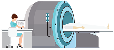
Today imaging in medicine has advanced to a stage that was inconceivable 100 years ago with growing medical imaging modalities including computed tomography, mammography, MRI, PET scan’s and more. The basic x-ray was a concept that spurred on research into other means of capturing images such as high frequency sound waves in ultrasound. To learn more about career options in the field of radiology, visit our radiology careers page or browse school programs offering radiology and ultrasound programs in your state.