
The field of radiology offers many opportunities for advancement and specialization. Many radiology degree programs offer certification in a particular area within the field. Radiology specializations will make you more competitive in the job market and will increase your earning potential.
The radiology specializations highlighted below are among the most common pursued by students in radiology degree programs. Request information from schools offering radiology programs for areas you find interesting.
The following radiology specializations conventionally fall under the umbrella of radiologic technologist. That is, most employees who perform the following jobs first became licensed Rad Tech’s, then obtained a certificate to perform the following radiology specializations:
The following is a list of common areas of radiology specializations under the umbrella of Ultrasound or Diagnostic Medical Sonographer (which is the preferred term in the medical imaging community). The most commonly known is Obstetric Sonography, which is the practice of monitoring the progress of a pregnancy; however, the field of Sonography entails much more than routine pregnancy check ups.
Bone densitometry, which is also called dual energy x-ray absorptiometry (DXA) is a form of x-ray technology that measures bone loss. DXA measures bone mineral density (BMD). It is often performed on the lower spine and hips to diagnose osteoporosis, which commonly effects women after menopause; however, bone loss is also found in men. Osteoporosis occurs when there is loss of calcium and other structural changes to the bones, which cause them to become thinner and more susceptible to fracturing.
An x-ray is a noninvasive medical test using a small dose of ionizing radiation to produce pictures of the inside of the body. The x-rays are transmitted through the patient to an image capturing device so that the physician can accurately make a diagnoses. There is the use of screen radiography, where the rays pass through the patient and then create an impression on film, which is developed chemically; however, more recently, the use of digital radiography has become used more often. X-Rays are the oldest type of medical image used and are commonly used to view broken bones, chest, as well as upper and lower GI tract.
CT Technicians (Computed Tomography techs) scan a particular part of the body using a large quantity of cross-sectional x-rays together with computer algorithms. An x-ray tube is used opposite a detector device in a ring shaped instrument; this rotates around a patient producing a computer generated cross-sectional image called a tomogram. CT Technician jobs also entail the use of substances which are intravenously inserted into the body in order to allow for greater contrast and accuracy while performing the scan. CT technicians work with more subtle variations than that of Radiographers and usually requires additional training. The recent advances in technology have lead to faster and more accurate scans, which has lead to higher demand for CT technicians and more CT technician jobs have resulted.
The use of low dose x-rays to produce images of the breast. The image is called a mammogram and is used to aid in the early detection and diagnosis of breast diseases and abnormalities such as cancer, lumps, etc. It plays a significant role in the early detection of breast cancer because it can show the beginnings of problems up to two years before a lump can be felt.
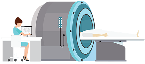
Images are produced using a combination of magnetic fields and radio waves.. MRIâs provide highly detailed three dimensional images of soft tissues. It provides very good contrast between the soft tissues than x-rays or computed tomography, which makes it especially useful in neurological (brain), musculoskeletal, cardiovascular, and oncological (cancer) imaging. MRIâs are a fairly new technology, with the first images captured in the mid 1970âs.
An imaging technique used to get real time images of the internal structures of a patient. It consists of an x-ray and a fluorescent screen and the patient is placed in between the two and an x-ray image intensifier and special video camera record the images on a monitor to view in real time. A radio-contrast such as a barium or an iodine is swallowed or injected into the body, which helps to delineate anatomical structures and the functioning of blood vessels.
Angiography is a technique in which x-ray technology is used to view the inside of blood vessels. It is typically used to view arteries, blood vessels, and heart chambers. A contrasting agent is injected into the blood vessels which the x-rays pick up and allow for viewing. Angiograms eliminate the bones and other anatomy, which allows you to view just the blood vessels.
Interventional radiology refers to non surgical treatments for medical conditions such as vascular diseases using radiologic technology. Examples include angioplasty, thrombolysis, atherectomy, embolization of bleeding vessels and occlusion of brain aneurysms. The procedures are performed with the guidance of x-rays, MRI’s, and ultrasound technology.
Cardiovascular interventional radiology consists of using imaging techniques for guidance while performing therapeutic procedures within the flow of blood to and from the heart. The pictures taken during the procedure are done using tiny instruments with small tubes, such as catheters, which provide a kind of road map that allow the Radiologist to navigate through areas of interest. One example, is angioplasty, which is used to widen a narrow blood vessel that may be blocked or to remove debris and plaque that is building up in the blood vessel. Another technique is the use of fluoroscopy to help guide the catheters. Interventional procedures can help to eliminate the need for surgical procedures.
Ultrasound technology is often used to gain access to veins being used for catheter placement to avoid possible complications such as bleeding during interventional procedures. Using ultrasound guidance, access is gained into veins while a small wire is inserted using x-ray guidance (fluoroscopy).
The use of sonography to evaluate the fetus and the female reproductive organs. This is a standard part of prenatal care in which the transducer is placed over the belly to capture images of the fetus or embryo. The image is examined for abnormalities or potential problems during the course of the pregnancy.
Cardiography is the use of ultrasound technology to view the heart, including the valve function and blood flow. The images can be viewed real time in three or even four dimensional. It is commonly used to monitor heart disease. It can show the size and shape of the heart as well as pumping capacity. It can help to detect coronary artery disease and a whole host of other heart related problems.
Use of sonography to image the musculoskeletal system of the body including tendons, ligaments, joints, nerves, muscles, and bone surfaces.
Use of sonography to obtain images of the female pelvic organs such as the uterus, fallopian tubes, cervix, ovaries, and bladder and to help in the diagnosis of problems associated with these organs.
The use of the doppler effect to enhance sonography procedures. In a nutshell, the doppler effect helps physicians understand the velocity and direction of blood flow. For example, the speed and direction of blood flowing through a heart valve can be examined using doppler technology, which is particularly useful in cardiovascular procedures.
PET (Positron Emission Tomography) is a nuclear imaging technique that produces a three dimensional image of the body by detecting gamma rays emitted by a radionuclide tracer which interacting with molecules. It is often performed in conjunction with CT x-ray scans to reproduce the images using computer algorithms on a scanner. The tracer is usually injected into a vein or swallowed and then gives off energy inside the body in the form of gamma rays. The energy is detected by the PET scanner and provides details on the functioning of organs and tissues. The use of PET with CT allows for a more precise information for diagnoses. It is commonly used to measure blood flow, oxygen use, glucose metabolism and to figure out how well an organ is functioning.
This is a specialty area within the field of nuclear medicine dedicated to compounding and dispensing radioactive materials for use in nuclear medicine procedures. As these procedures become more commonplace, specialists are needed to prepare and label the radionuclides. As a Nuclear Pharmacist, you will work in a “drugstore” for the nuclear medicine department. They are responsible for obtaining the desired radioactive material and dispensing it behind leaded glass shielding.
Radiation therapy is the use of high energy radiation to treat cancer. The goal of radiation therapy is to destroy the cancer cells’ ability to reproduce, which allows the body to naturally get rid of the remaining cells. The use of radiation therapy can prevent the need to remove entire organs such as the breast or bladder during a surgical procedure.